BEMER Electromagnetic Field Therapy Reduces Cancer Cell Radioresistance by Enhanced ROS Formation and Induced DNA Damage
Abstract
Each year more than 450,000 Germans are diagnosed with cancer and receive standard therapies including surgery, chemotherapy, and radiotherapy. Due to intrinsic and acquired resistance, alternative therapeutic support is considered, including non-invasive complementary approaches.
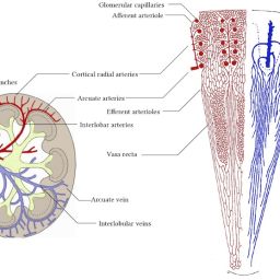
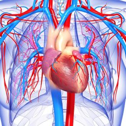
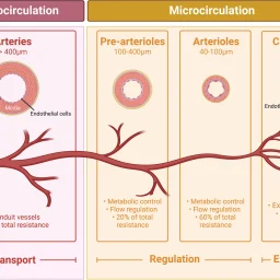
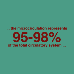
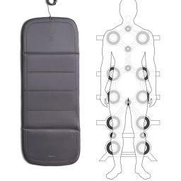
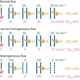
BEMER therapy, a physical vascular therapy applying low-frequency pulsed EMF, normalizes microcirculation and has been investigated for its impact on cancer cell survival during radiotherapy.
Using three-dimensional cell culture models from lung, head and neck, colorectal, and pancreatic cancer, BEMER treatment enhanced radiosensitization, increased ROS levels, and elevated DNA double strand breaks (DSBs). However, it did not sensitize cells to chemotherapy or Cetuximab.
Study Findings
BEMER therapy decreases radioresistance dependent on signal intensity
BEMER therapy applied at 2.7–35 μT 1 hour after 6-Gy X-ray irradiation increased DNA double strand breaks in A549 and UTSCC15 cells, suggesting a link between signal intensity and radiosensitization.

BEMER therapy increases ROS levels leading to radiosensitization
ROS scavengers (sodium pyruvate, MnTBAP, Carboxy-PTIO) were used to test ROS involvement. MnTBAP and Carboxy-PTIO reduced DSB numbers and abrogated radiosensitization, confirming ROS-mediated effects.

Original Study
For the full version of the study, visit: https://www.ncbi.nlm.nih.gov/pmc/articles/PMC5154536/
Citation: PLoS One. 2016; 11(12): e0167931. doi: 10.1371/journal.pone.0167931 – PMCID: PMC5154536 – PMID: 27959944
Physical Vascular Therapy in Practice – Scientific Evidence and Therapeutic Applications